
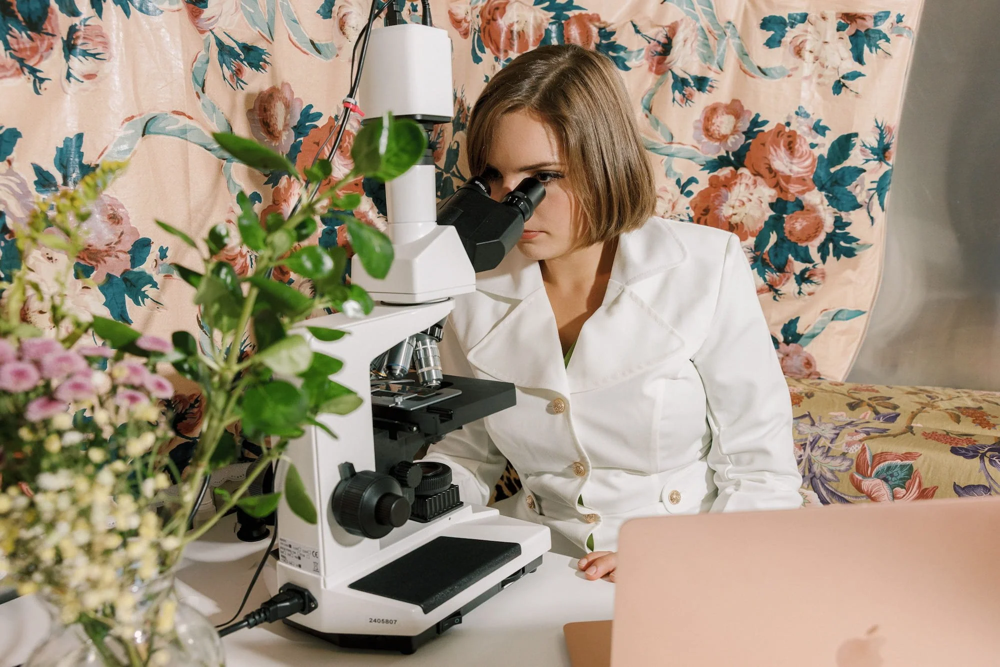
Live Blood Microscopy
A Window into Your Body's Inner Terrain
A single drop of your blood, observed live and unaltered under a high-powered microscope, reveals what standard lab panels often miss. It is the difference between a still photo and a film — you see your body’s terrain in motion.
Methodology informed by the work of Dr. Okker & NeoGenesis Systems.
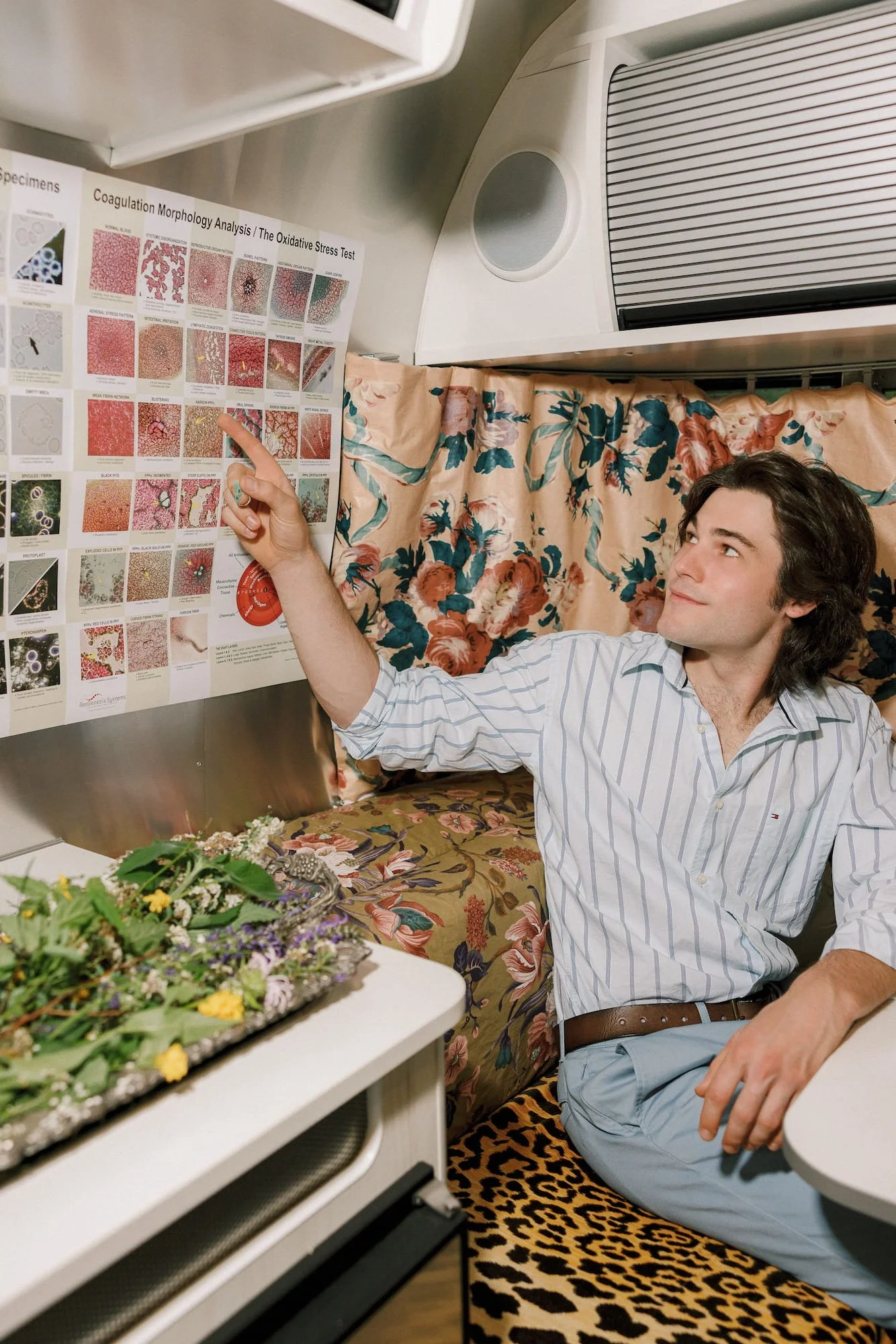
THE PRACTICE
What It Is
Live blood microscopy (sometimes called dark-field or live-cell analysis) uses a single drop of capillary blood from the fingertip, viewed in real time on a high-resolution screen. The blood is fresh, living, and unstained — so what you see is exactly how your blood is behaving inside the body, not a preserved snapshot.
Unlike a standard lab draw — which measures quantities of cells and markers — live blood microscopy is qualitative and observational. We watch how the cells move, how the plasma flows, what is in suspension, and what story the terrain is telling.
Our Lineage
The methodology we use here is rooted in the foundational work of Dr. Okker and the NeoGenesis Systems framework — a school of live and dark-field microscopy focused on terrain assessment, biological-medicine principles, and the relationship between blood quality and whole-body vitality. What you see in your session draws directly from that lineage.
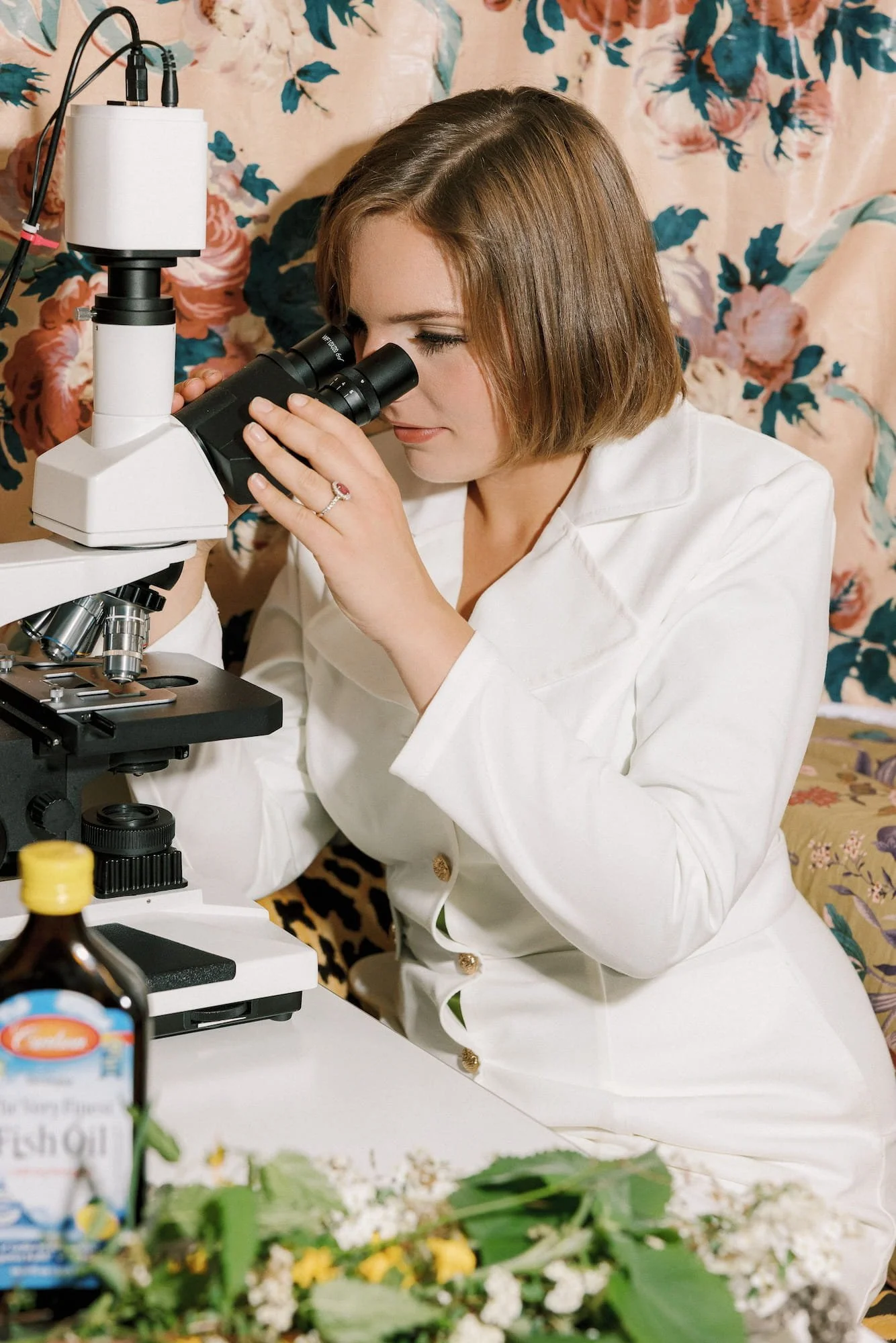
IN SESSION
How It Works
01
Single Drop
A small drop of blood is taken from the fingertip and placed on a slide.
02
Live Viewing
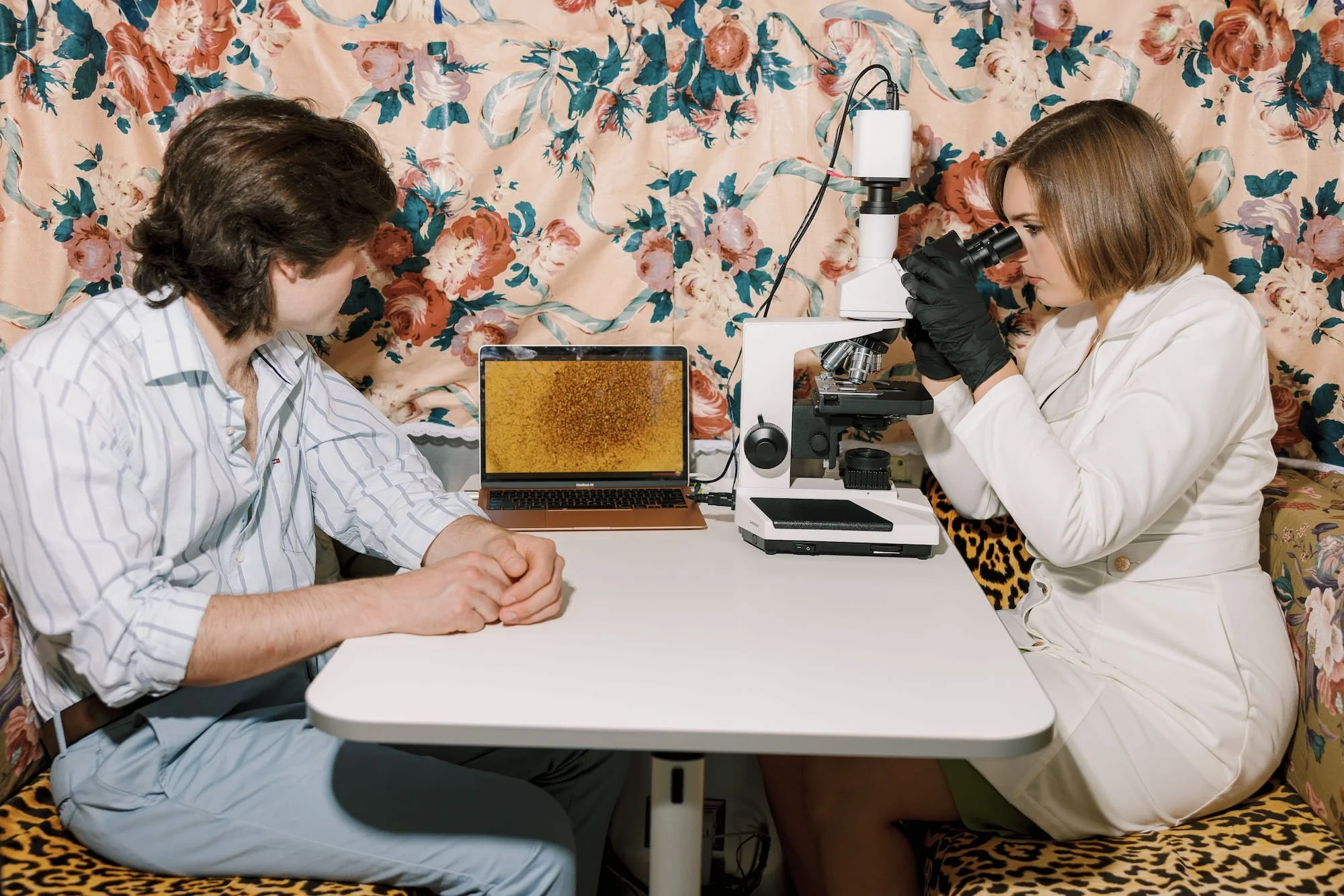
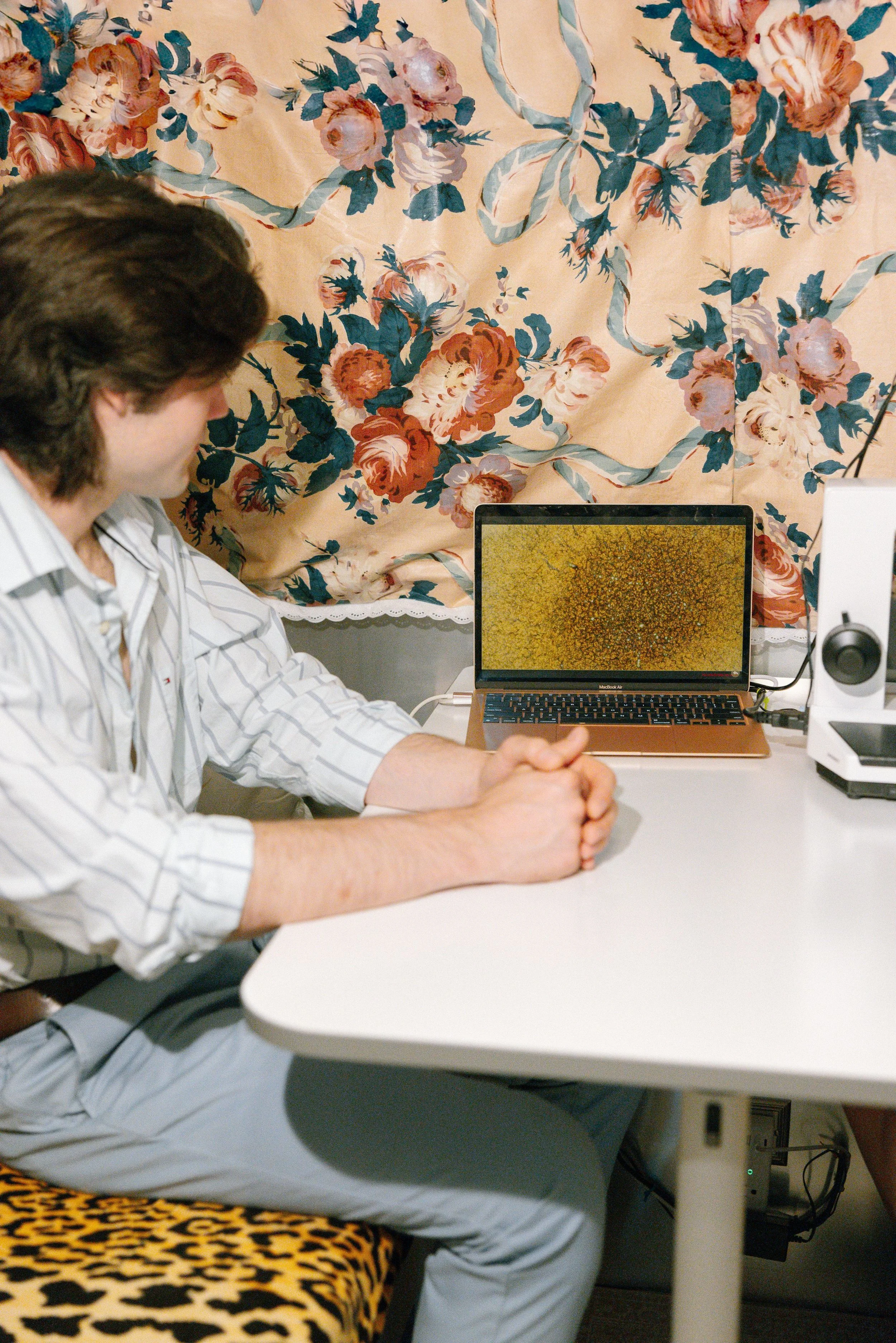
The blood is observed in real time on a high-resolution screen beside you.
03
Walk-Through
We narrate what we are seeing — cell shape, plasma quality, terrain.
04
Personalized Plan
Findings inform a clear set of next steps tailored to you.

AI-Powered Analysis
A MODERN EDGE
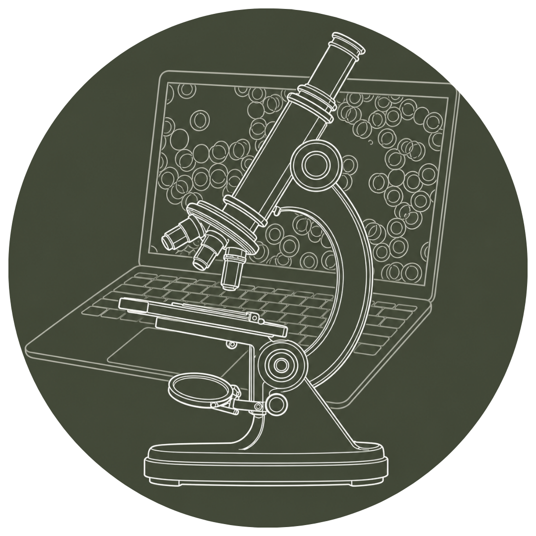
Our practice is equipped with proprietary AI analysis technology developed by Dr. Okker's Live Blood Online — a tool trained on years of clinical imagery that pairs the trained human eye with machine pattern recognition. The result: a more thorough, consistent, and reproducible read of every drop.
Pattern Recognition
AI identifies subtle cell shapes, plasma patterns, and anomalies the eye can miss in real time.

Reference Library
Your images are compared to thousands of trained samples to surface known indicators and trends.

Tracked Over Time
Sessions are saved and compared visit-to-visit so progress is measured, not guessed.
Practitioner-Led
The AI is a tool, not a verdict. Every observation is interpreted alongside your story and goals.
Inside the Drop —
What We Can See
Each drop holds a remarkable amount of information. There are over 50 abnormalities that can be seen. A few of the patterns and signals we look for:
Red Blood Cells
Shape, Size & Mobility
Healthy red cells move freely and are uniform in shape. Stacking (rouleaux), distortion, or clumping points to oxygenation, hydration, and digestion patterns.
Microorganisms
Yeast, Bacteria & Parasites
Live blood microscopy can reveal yeast/candida overgrowth, fungal forms, bacterial activity, and parasitic indicators that do not always show on standard panels.
Digestion
Gut & Absorption Signals
Undigested fats, food particles, and certain plasma patterns suggest leaky gut, low stomach acid, or compromised absorption.
Circulation
Flow & Oxygen
Sluggish blood, sticky cells, and poor cell-to-cell separation reflect circulation, oxygen delivery, and overall vitality.
Plasma
Terrain & Clarity
A clean plasma background means the body is processing and clearing well. Cloudy plasma, fibrin webs, or protein linkage suggest inflammation, liver stress, or congested terrain.
Oxidative Stress
Free Radical Activity
Crystalline formations, plaque, and damaged cells reflect oxidative load — a key driver of aging and chronic disease.
Nutrition
Deficiency Clues
Cell shape and size offer clues to common deficiencies — iron, B12, folate, essential fats, and protein status.
White Blood Cells
Immune Activity
We watch how white cells move and respond — a window into immune vitality, current infections, or chronic immune burden.
Heavy Metals & Toxins
Tell-Tale Patterns
Specific cell distortions and crystalline structures hint at heavy metal load and environmental toxin burden.
Hydration
Fluid & Electrolytes
How the cells move and space themselves on the slide is one of the fastest reads on hydration status.
"For many members, seeing their own blood for the first time changes everything. It transforms abstract health conversations into something tangible, helping them better understand their body and feel more engaged in their wellness journey."
The Benefits
WHY IT MATTERS
01 A Real-Time View of Your Body - No waiting weeks for lab results — you see what is happening in your body the moment it is happening.
02 A Starting Baseline You Can Return To - Your first session establishes a visual baseline. Future sessions show progress, regression, or response to specific protocols.
03 A Personalized Roadmap - What we observe directly shapes your supplement, food, hydration, and lifestyle plan — tailored to your terrain, not a generic protocol.
04 Early Signals Before Symptoms - Many patterns — oxidative stress, sluggish liver, immune burden — show up in the blood long before you feel them in the body.
05 Powerful Motivation - There is nothing like seeing your own blood improve to keep you committed to the work. Visual feedback is one of the most underrated tools in healing.
06 An Education in Your Own Body - You leave knowing things about how your body works that you have never known before — and with a way to keep tracking it.

An Honest Note
Live blood microscopy is an educational and observational practice — not a diagnostic medical test. It complements standard lab work and conventional medicine; it does not replace either. What it offers is something unique: a real-time, qualitative view of your terrain that informs better questions, better choices, and a clearer path forward.
See Your Body in Motion
Whether you are curious, healing, or simply optimizing — this is one of the most direct, intimate views of your own biology you can have. Bring your questions. Bring your goals. We will show you what is there.
Preparing For Your Session
Come as you are — rested, hydrated, and curious. A few small adjustments make a meaningful difference in what we are able to see together under the microscope:
-
Please arrive having not eaten for at least three hours before your appointment for the clearest reading. Water is welcome and encouraged.
-
Dehydration changes how your blood moves and presents. Drink water steadily in the hours leading up to your visit.
-
Hold off on your supplements the morning of your test so we can capture your true baseline.
-
If possible, schedule your visit during your follicular phase (roughly the week after your period begins) for the most representative picture.
-
If you are actively detoxing, your blood may temporarily look more congested as toxins mobilize. This is a sign of progress, not a setback.
-
Each of these factors influences what we see under the microscope. The more honestly you arrive, the more useful the read. We'll take it from there.
Experience Live Blood Microscopy at Floral Botanical Medicine
Located in Rumford, Rhode Island, our custom Airstream wellness studio offers a calm, private setting where you'll experience Live Blood Microscopy alongside personalized education and botanical wellness guidance.
Membership is free and provides access to this unique educational experience, along with our full range of member wellness services.